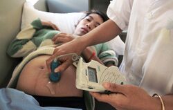
Prenatal Ultrasound
(medical procedure) | |
|---|---|
 | |
| A taken-for-granted component of modern maternity care..but critics say it may cause permanent and heritable damage for ten generations and beyond. |
Prenatal ultrasound (obstetric sonography) is the name for a number of tests done during the pregnancy of a woman, using ultrasound to visualize hidden body structures. Prenatal ultrasound is a taken-for-granted component of modern maternity care, to such an extent that most obstetrician-gynecologists find it impossible to practice their profession without it. [1][2]
Contents
Official narrative
The World Health Organization states:"Diagnostic ultrasound is recognized as a safe, effective, and highly flexible imaging modality capable of providing clinically relevant information about most parts of the body in a rapid and cost-effective fashion".[3]
American women routinely undergo four to five ultrasounds per pregnancy.[2]
Cellular damage
Ultrasound technology is not just sound waves but is based on non-ionizing radiation[4]. Other examples of man-made non-ionizing radiation include cell phones, cell towers, cordless phones, Wi-Fi and more. Although ionizing radiation (for example X-rays) has the reputation of being more powerful, non-ionizing radiation is definitely capable of producing biological effects—including altering and damaging cells.[2]
According to author Jeanice Barcelo[5], the technology causes far-reaching damage. Describing a series of studies published in the late 1970s[6] and early 1980s[7], she notes that “a single exposure to ultrasound produced cellular and DNA damage similar to 250 chest x rays”—and “[d]amage was permanent and heritable for ten generations and beyond.[8]” Forms of damage included “DNA shearing, single and double strand breaks, chromosome rearrangements and DNA uncoiling, deformities and mutations in offspring, as well as the complete deactivation of genetic material within sonicated cells.”[9]
A study performed in Sweden in 2001[10] has shown that subtle effects of neurological damage linked to ultrasound were implicated by an increased incidence in left-handedness in boys (a marker for brain problems when not hereditary) and speech delays.[11][12] The above findings, however, were not confirmed in a follow-up study.[13]
The technology and its applications have continually expanded. Besides the huge increase in allowable acoustic output in the early 1990s, the following changes have made the field of prenatal ultrasound riskier[14]:
- The number of ultrasound scans conducted during each pregnancy has increased, with women often receiving two or more scans even in low-risk situations. Women in "high-risk" situations may receive many more scans—which, ironically, may raise their risk.[14]
- The range of time within an embryo or fetus's development when ultrasound is performed has extended to very early in the first trimester and continues into the third trimester, right up to delivery. Fetal heart monitors that are used prior to delivery—sometimes for hours—have not been shown to reduce neurological problems and may increase them.[14]
- The development of the vaginal probe, which positions the beam of sound much closer to the embryo or fetus, may put it at higher risk. [14]
- The use of Doppler ultrasound, which is used to study blood flow or to monitor the baby's heartbeat, has increased. According to the 2006 Cochrane Database of Systematic Reviews, "routine Doppler ultrasound in pregnancy does not have health benefits for women or babies and may do some harm."[14]
Selective abortion as population reduction tool
In a 1967 memo, the Population Council discussed various methods to reduce the world population. One of the priorities was "development of more powerful institutions to deal with population problems", where one of the priorities was "augmented research efforts leading to improved contraceptive technologies or practical methods of determining the sex of an unborn child so that parents could be assured of a son".[15]
In the mid-1960s, the leading American embryologist and biochemist, Sheldon Segal, working the Population Council, showed doctors at India's top medical school AIIMS how to test human cells for sex chromatins that indicate whether a person is female, a method that was the precursor to foetal sex determination. In India, the early sex selective abortions were performed openly at government hospitals. Western money (Ford Foundation, Rockefellers..) was used to set up an extensive network of family planning advisers and doctors that encouraged women to opt for amniocentesis, the precursor to ultrasound method.[16]
From the 1970s onwards, medical diagnostic testing using ultrasounds became commonly available to determine the sex of a fetus during pregnancy. The technology made selective abortion of female fetuses possible, a practice that is most common where male children are valued over female children, especially in parts of East Asia and South Asia (particularly in countries such as China, India and Pakistan)[17], this despite legal bans on the practice.[18]
Since sex determination technology became available, India is estimated to have about 63 million fewer women, leading to a skewed ratio of men to women – as of 2020 between 900-930 females per 1,000 males.[19][20]
References
- ↑ https://www.ncbi.nlm.nih.gov/pubmed/24176148
- ↑ Jump up to: a b c https://childrenshealthdefense.org/child-health-topics/known-culprits/ultrasound/prenatal-ultrasound-not-so-sound-after-all/
- ↑ http://whqlibdoc.who.int/trs/WHO_TRS_875.pdf
- ↑ https://humanhealth.iaea.org/HHW/MedicalPhysics/DiagnosticRadiology/DosimetryInstrumentationandCalibration/NonIR/index.html
- ↑ https://birthofanewearth.com/2019/04/the-dark-side-of-prenatal-ultrasound/
- ↑ https://pubmed.ncbi.nlm.nih.gov/424580/
- ↑ https://www.ncbi.nlm.nih.gov/pubmed/7455124
- ↑ https://www.electricsense.com/ultrasound-pregnancy-risks/
- ↑ https://www.electricsense.com/ultrasound-pregnancy-risks/
- ↑ https://doi.org/10.1097%2F00001648-200111000-00007
- ↑ https://doi.org/10.1136%2Fbmj.307.6897.159
- ↑ https://doi.org/10.1016%2FS0378-3782%2897%2900097-2
- ↑ https://doi.org/10.1002%2Fuog.8962
- ↑ Jump up to: a b c d e https://www.midwiferytoday.com/mt-articles/questions-prenatal-ultrasound/
- ↑ quoted in the ARTE documentary Bloß keine Tocher/ Un monde sans femmes at time 39:17
- ↑ https://www.bbc.com/news/14213136
- ↑ https://doi.org/10.1080%2F00324720308069
- ↑ See https://en.wikipedia.org/wiki/Pre-Conception_and_Pre-Natal_Diagnostic_Techniques_Act,_1994
- ↑ https://pha.berkeley.edu/2021/04/10/un-natural-selection-female-feticide-in-india/
- ↑ https://www.theguardian.com/global-development/2020/aug/21/selective-abortion-in-india-could-lead-to-68m-fewer-girls-being-born-by-2030
